




MicrUs
Ecografo Ultra-Portatile
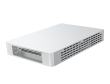
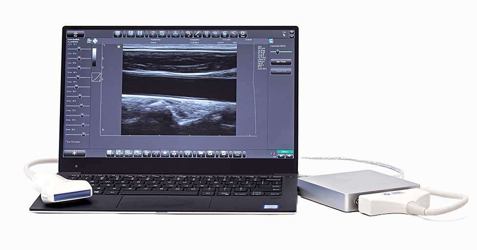
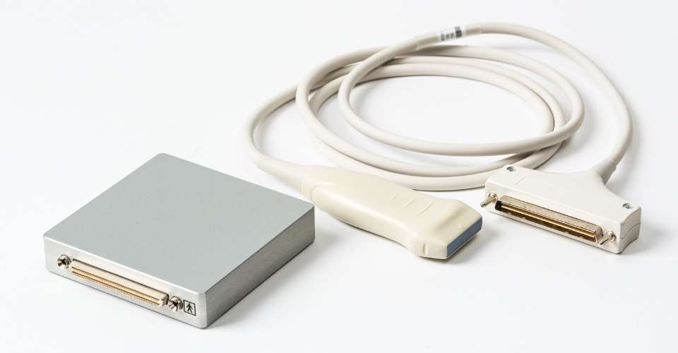
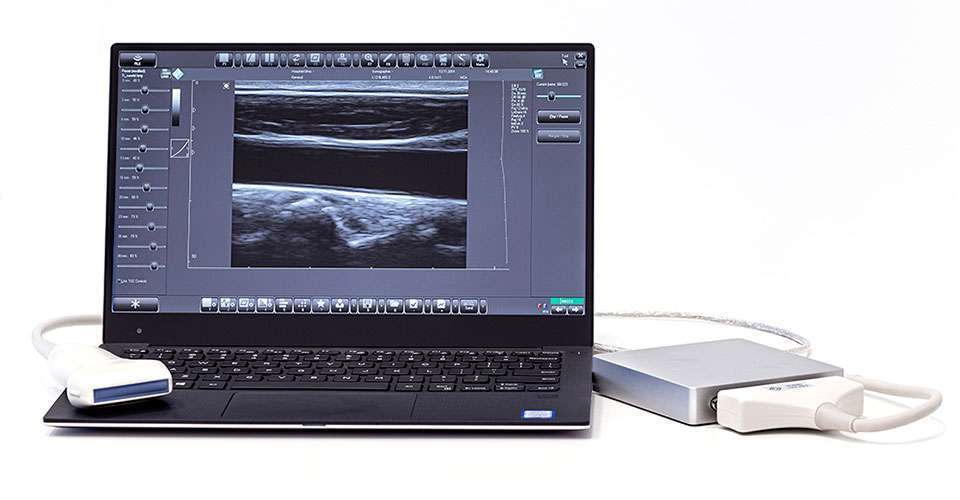
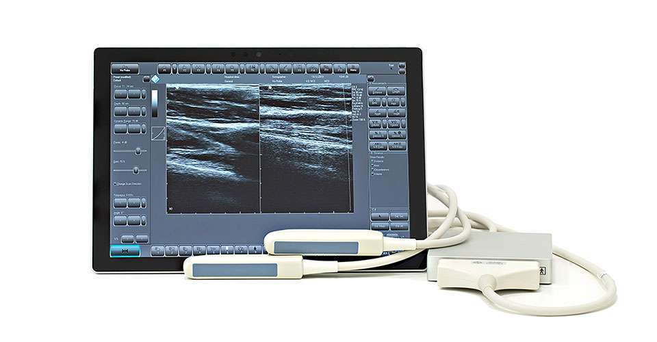
Il MicrUs EXT-1H è un ecografo tascabile piccolo e compatto che fornisce immagini ad alta definizione con il tuo smartphone, tablet o PC e supporta un’ampia gamma di sonde.
Ecografo MicrUs
in breve
Ultraleggero e Compatto
Uno scanner ecografico tascabile di nuova generazione per il point of care, senza ventola e alimentato via USB.
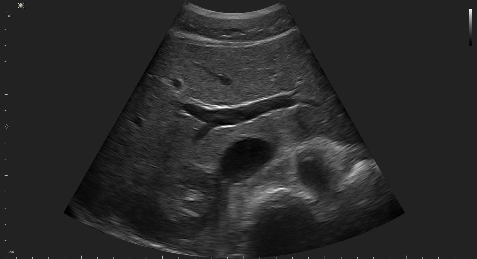
Immagini di Qualità
Eccellenza Software
Il software Echo Wave II è stato progettato per offrire una suite completa di strumenti diagnostici ed è aggiornabile gratuitamente.
SDK & Strumenti di Ricerca
Il MicrUs offre funzioni opzionali per la ricerca e sviluppo come il kit di sviluppo software ed il modulo I/O per la sincronizzazione.
Ecografo Ultraportatile USB
Interfaccia e Alimentazione USB
Progettato per offrire massima portabilità e praticità, eliminando la necessità di un alimentatore esterno. Questo scanner ecografico tascabile si alimenta direttamente tramite porta USB, riducendo ingombri e facilitando l’uso in qualsiasi ambiente clinico.
Ampia gamma di trasduttori intercambiabili
Tecnologia avanzata di riconoscimento delle sonde per collegare e scollegare vari trasduttori in pochi secondi. Questa funzionalità consente una rapida transizione tra diverse applicazioni cliniche, migliorando l’efficienza durante le procedure diagnostiche.

Software Installabile su più dispositivi
Software versatile che può essere installato su più dispositivi Windows e Android, senza ulteriori costi. Gli aggiornamenti del software sono gratuiti, contribuendo a ridurre i costi di manutenzione e garantire sempre accesso alle ultime funzionalità e miglioramenti.
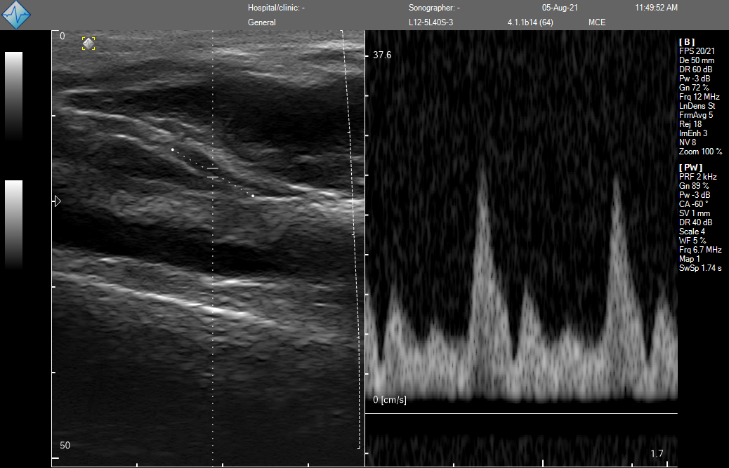
Pulsed Wave Doppler, Spatial Compound, B-steer, Wide View, Speckle Reduction Avanzati
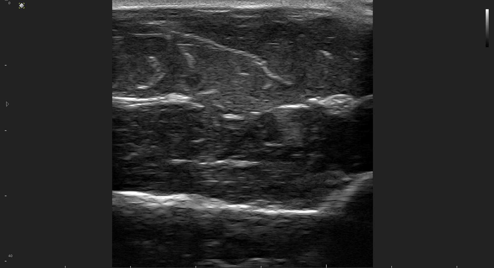
Screening ecografici di qualità in modalità B, M, B+M, 4B, PW Doppler, PW Doppler+B (duplex) con Speckle Reduction Processing, Trapezoid e Spatial compound imaging.
MicrUs in azione.
Ottieni dettagli incredibili per la tua specialità. Con la potente tecnologia e le schede di circuito attentamente progettate del MicrUs Ext-1H avrai a disposizione la potenza di un ecografo ad alte prestazioni.



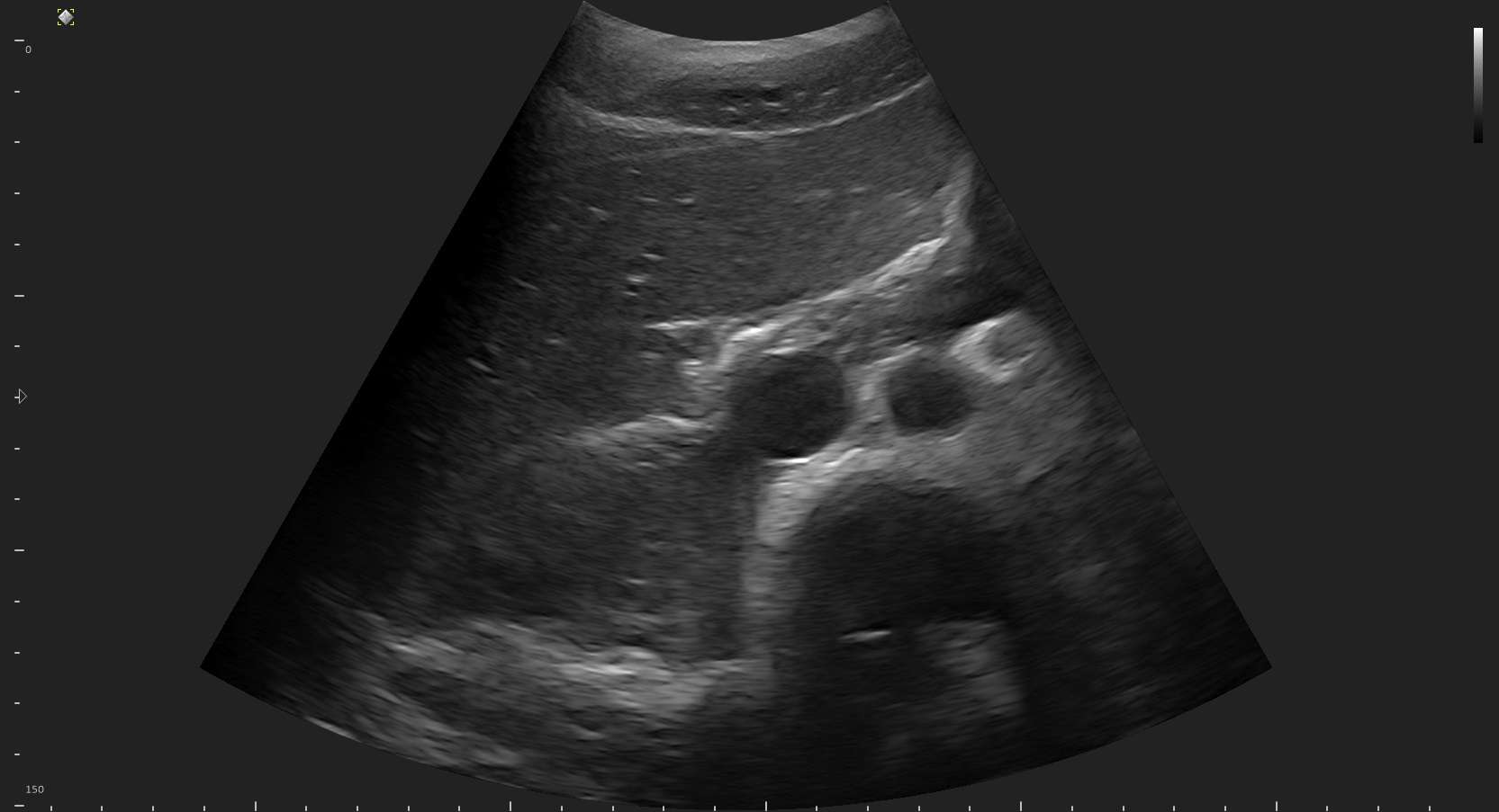
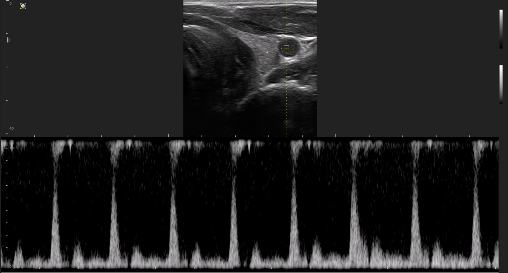

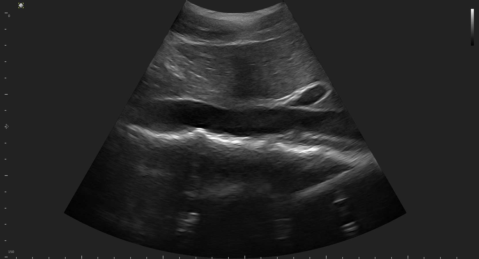


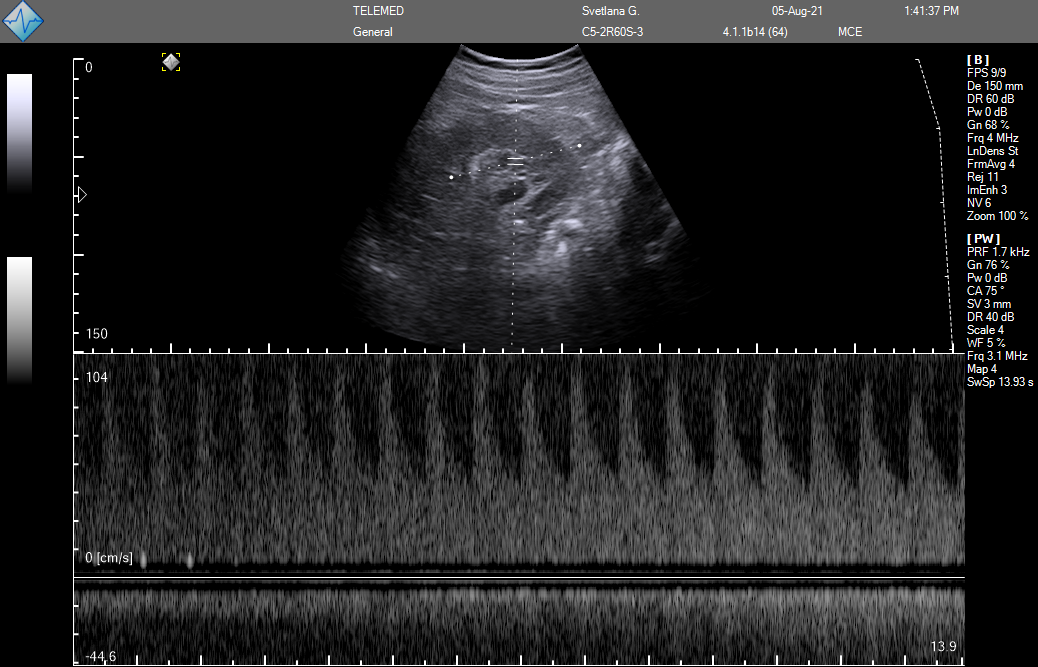


Modelli
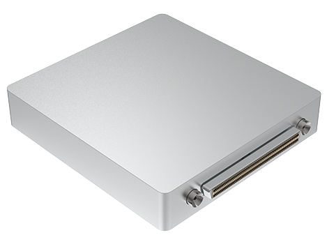
MicrUs EXT-1H
Ecografo Portatile Multidisciplinare
Caratteristiche
Connettori per sonde
Una porta per sonde
Supporto al prodotto
Garanzia
MicrUs: 2 anni – Sonde: 1 anno
DICOM
Modalità di scansione
B-Mode / M-Mode
Pulsed-Wave Doppler
Software di scansione
Echo Wave II – Ultrasound Software
Echo Wave II – Ultrasound Software Touch GUI
Echo Wave A – Android Ultrasound App
Strumenti per ricerca e sviluppo
SDK – Software Developement Kit
Richiede NDA
Real-time Imaging for the Research
Modulo I/O
Optionale*
Certificazioni
CE
FDA
Modello
Porte per sonde: Una
- Frequenza: da 2.0 a 15.0 MHz
- Trasduttori compatibili: Lineare, Convex, Microconvex
- Applicazioni: Vascolare, Piccole parti, MSK, Pediatria, Cardiologia, Accessi Vascolari, OB/GYN, Transrettale, Transvaginale
- Sistema operativo: Windows/Android
- Modalità di scansione: B-mode, M-mode, Pulsed Wave Doppler
- DICOM: ✔️
- Due anni di garanzia: ✔️
- Supporto al prodotto: ✔️
Scopri l’ampia gamma di sonde disponibili.
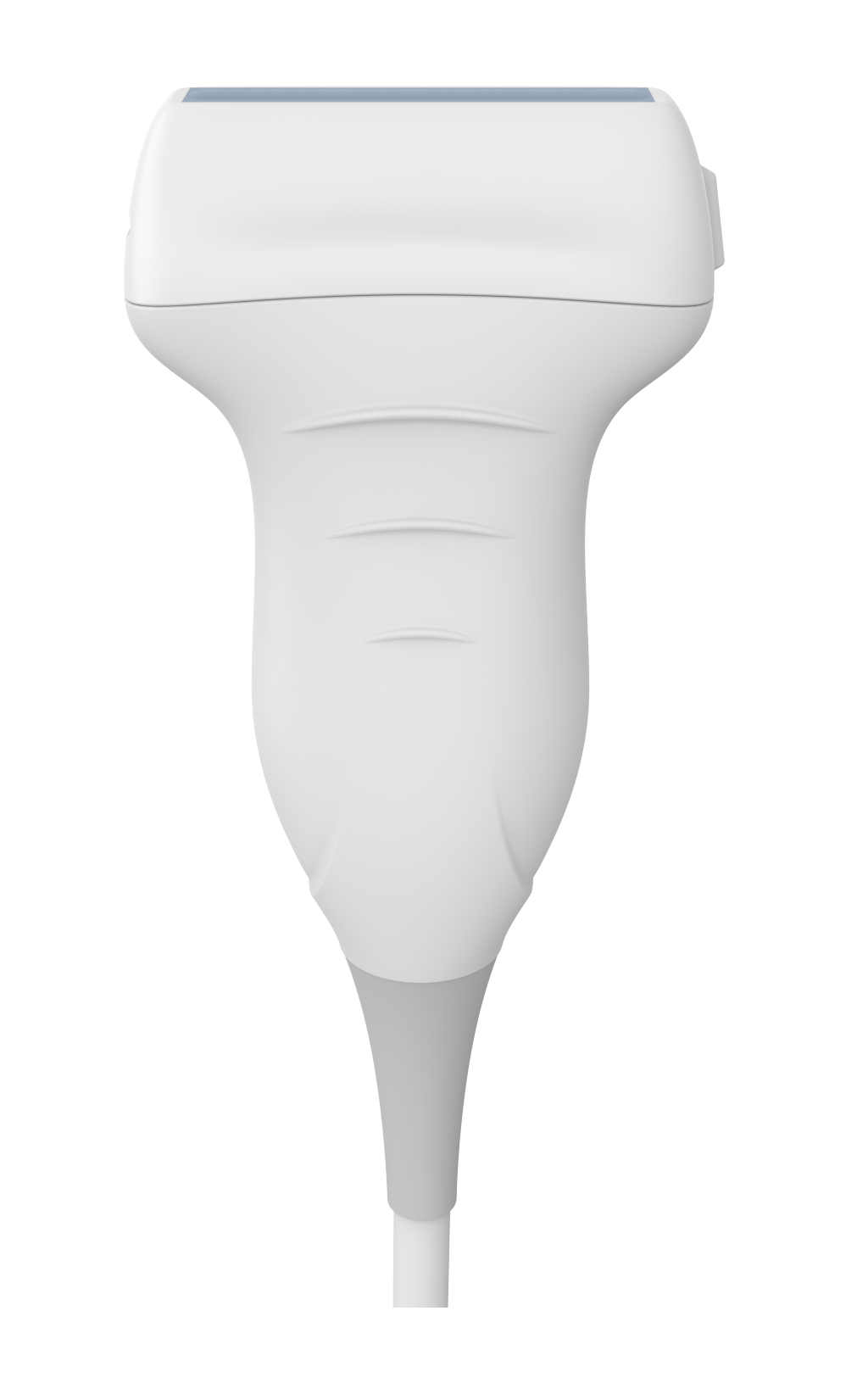
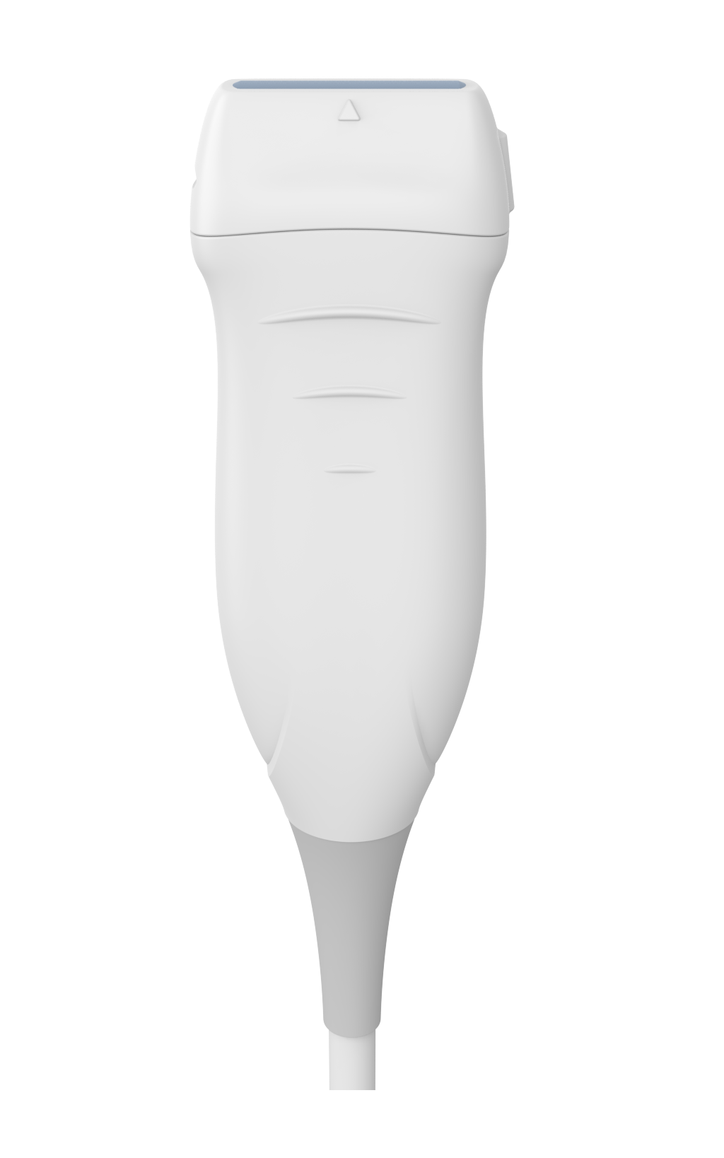
L12-5L40S-3
Type: Linear
Frequency range: 5.0 – 12.0 MHz
Field Of View: 38 mm
Applications: Vascular, Small Parts, MSK/Neurol., Pediatrics, Vascular Access
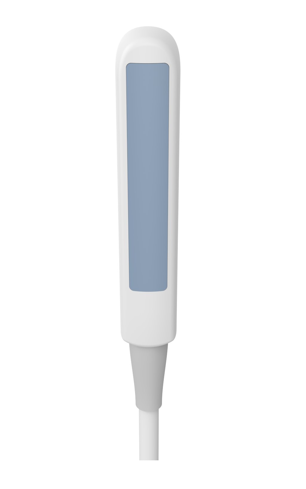
L15-6L25S-3
Type: Linear
Frequency range: 6.0 – 15.0 MHz
Field Of View: 24 mm
Applications: Vascular, Small Parts, MSK/Neurol., Pediatrics, Vascular Access
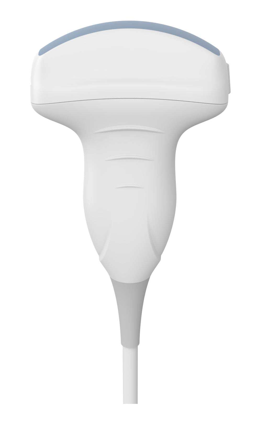
LV8-4L65S-3
Type: Linear
Frequency range: 4.0 – 8.0 MHz
Field Of View: 65 mm
Applications: MSK/Neurol.
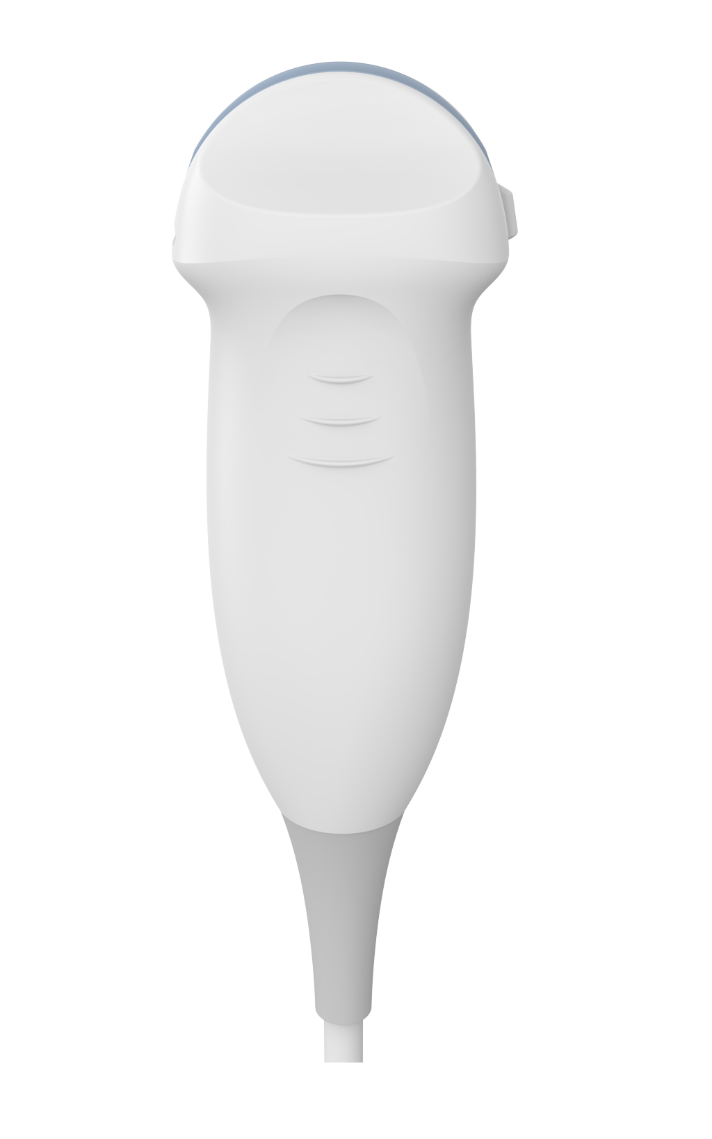
C5-2R60S-3
Type: Convex
Frequency range: 2.0 – 5.0 MHz
Field Of View: 60°
Applications: Abdomen, OB/GYN, Pediatrics
MC8-4R20S-3
Type: Micro Convex
Frequency range: 4.0 – 8.0 MHz
Field Of View: 91.7 mm
Applications: Small Parts, MSK/Neurol, Pediatrics
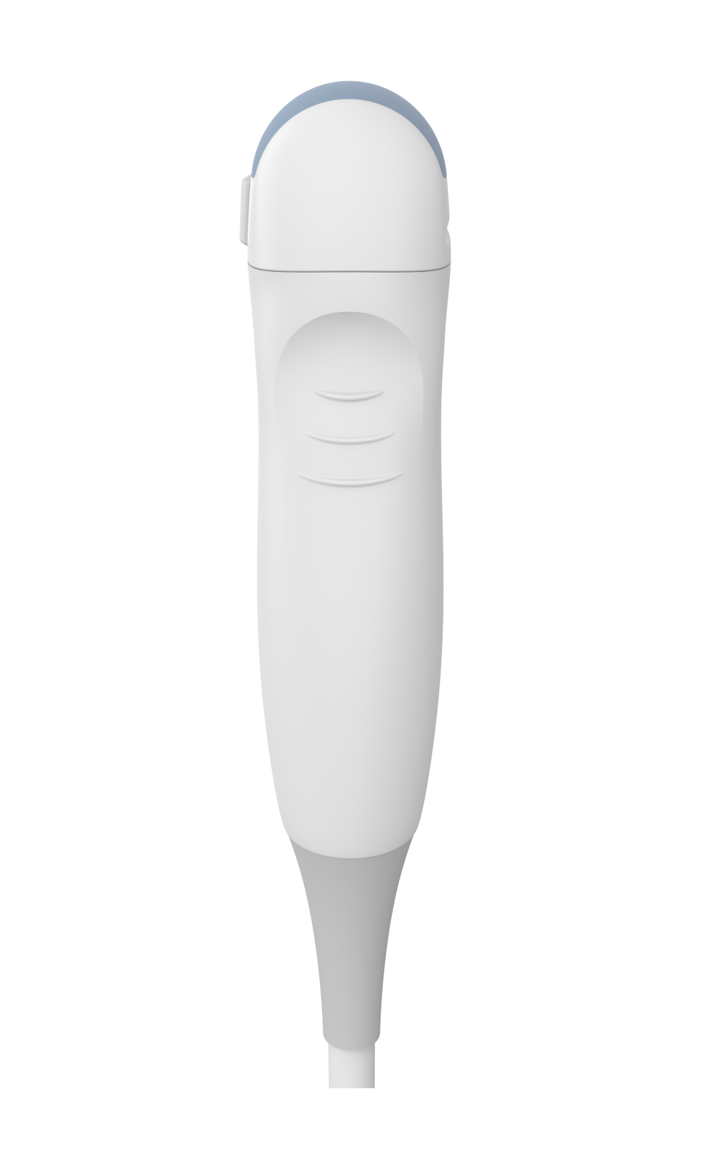
MC10-5R1-S-3
Type: Micro Convex
Frequency range: 5.0 – 10.0 MHz
Field Of View: 156°
Applications: Small Parts MSK/Neurol., Pediatrics, Cardiology
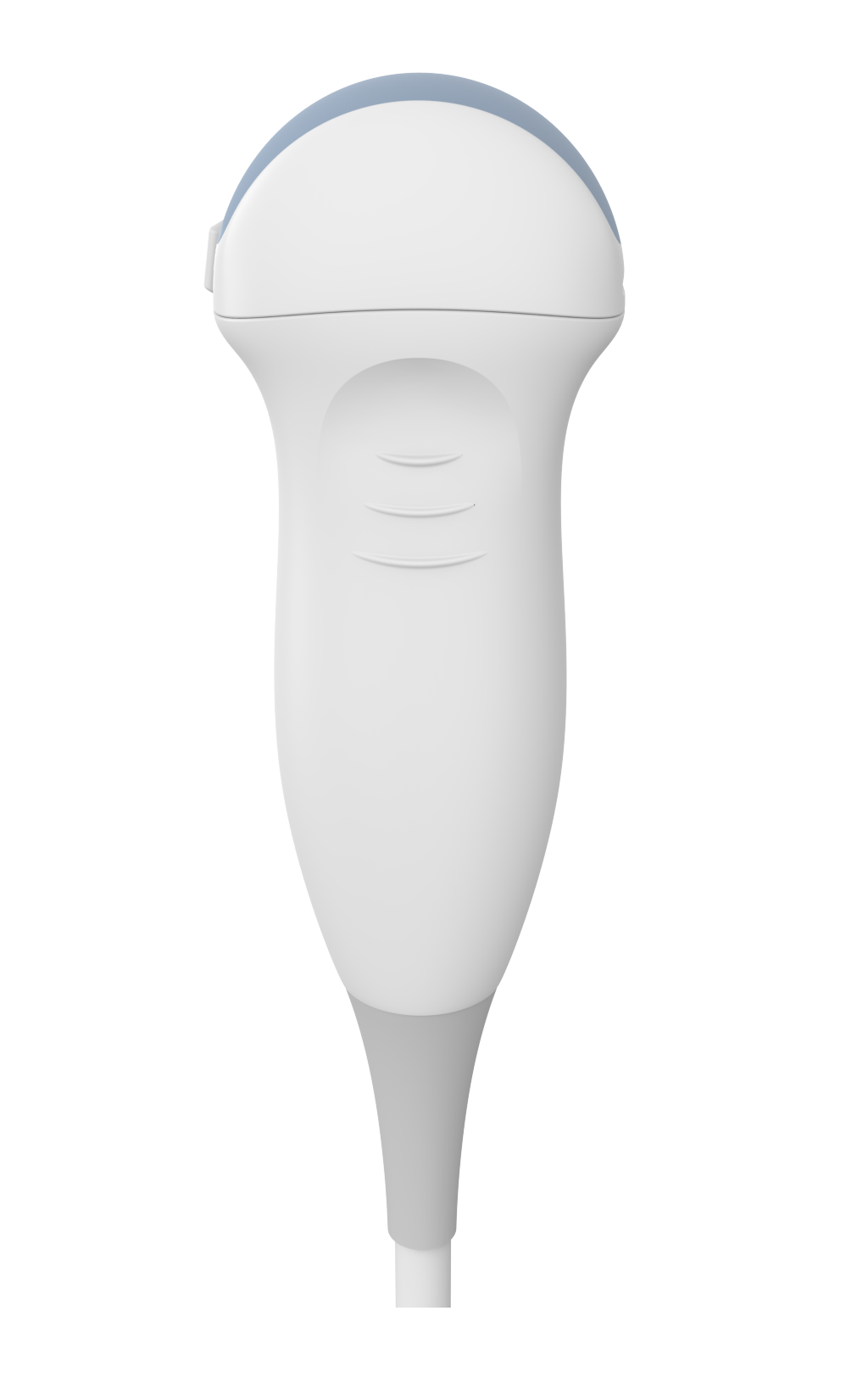
MC4-2R20S-3
Type: Micro Convex
Frequency range: 2.0 – 4.0 MHz
Field Of View: 104°
Applications: Abdomen, Pediatrics
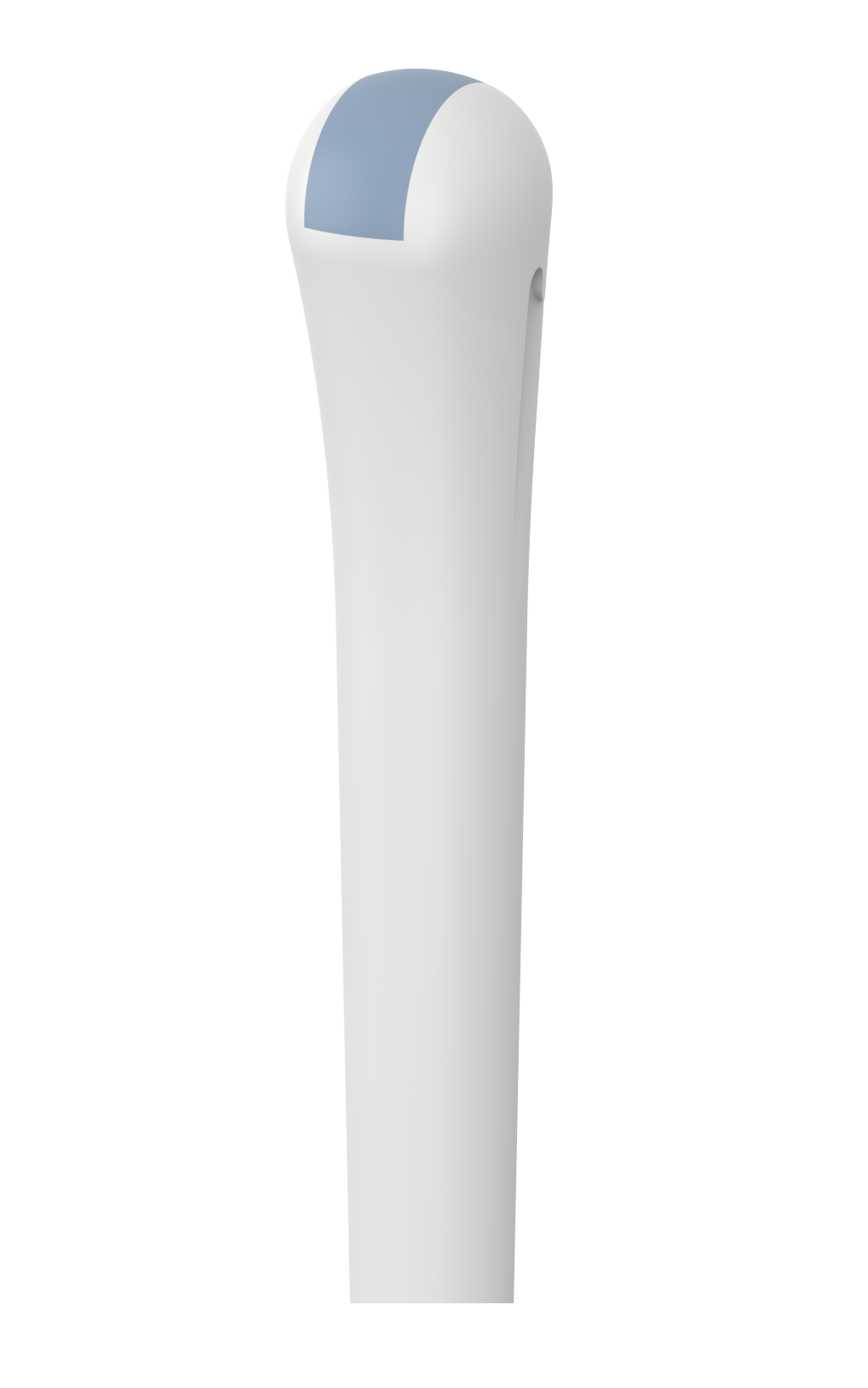
MCV9-5R10S-3
Type: End-Fire / Micro Convex
Frequency range: 5.0 – 9.0 MHz
Field Of View: 156°
Applications: Transrectal, Transvaginal
Strumenti di ricerca intuitivi, modulari e pronti all’uso per un tempo di configurazione ottimizzato.
Mettiamo a disposizione strumenti intuitivi per gli ambienti Python, MATLAB e LabView, arricchiti da una documentazione dettagliata e istruzioni comprensibili, permettendo così una notevole riduzione della curva di apprendimento.
DLL per MATLAB e Python
Libreria di collegamento dinamico (DLL) che consente di richiamare le funzioni dell’SDK e di eseguire l’acquisizione di dati e immagini in tempo reale da altri programmi come MATLAB, Python e Labview.
SDK – Software Development Kit
L’SDK di sviluppo software di scansione a ultrasuoni è una libreria di programmazione ad alto livello che consente lo sviluppo di software di scansione a ultrasuoni per tutti i sistemi ecografici TELEMED su Windows, mentre su Android è disponibile per i sistemi MicrUs e MicrUs Pro.
*Il pacchetto SDK completo è disponibile gratuitamente per partner OEM e sviluppatori dopo la firma dell’NDA con il produttore.
Modulo I/O
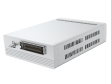
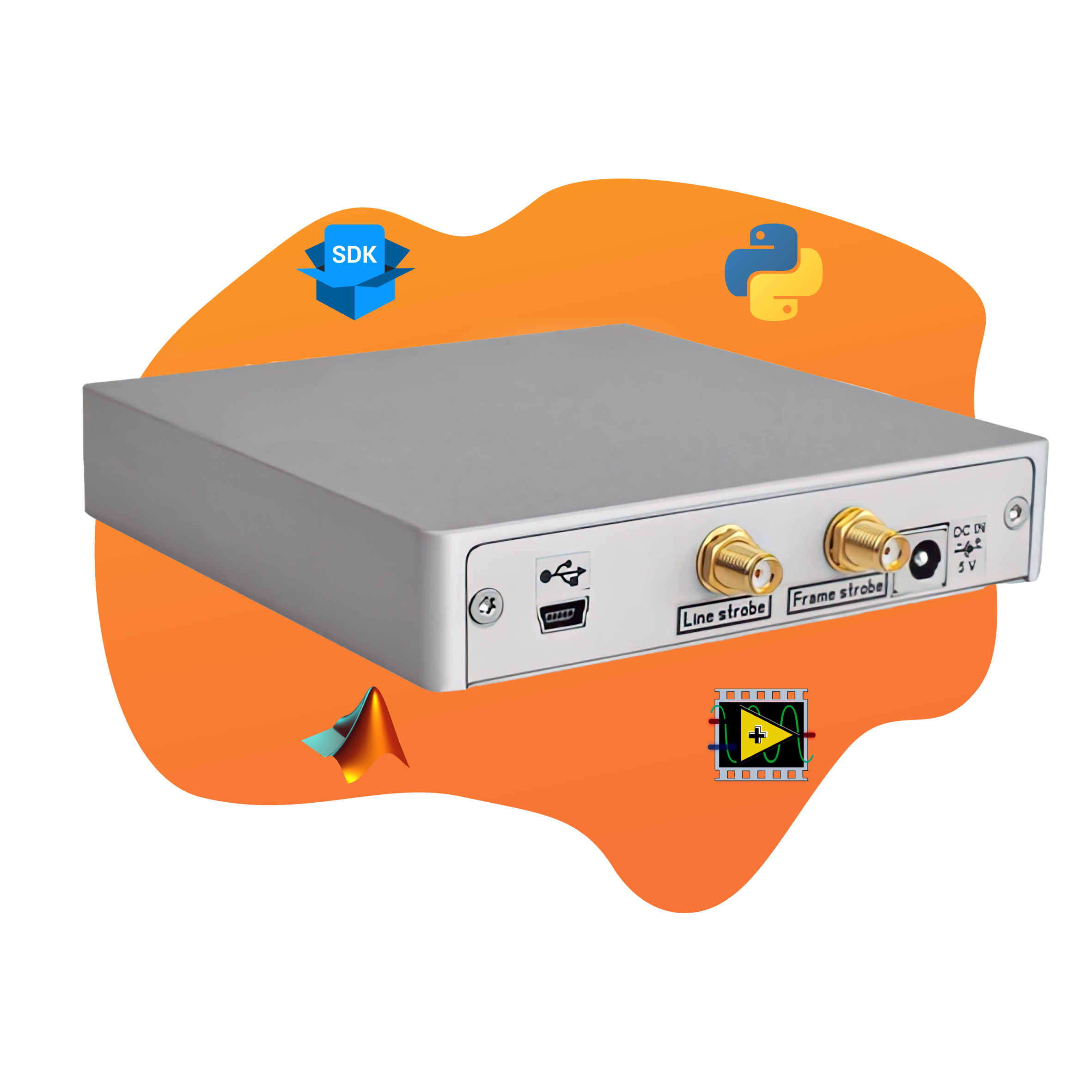
Il MicrUs può essere dotato di connettori aggiuntivi per la sincronizzazione del sistema con altre apparecchiature.
*Il modulo I/O è una feature opzionale.
Specifiche Tecniche
Models
MicrUs EXT-1H: one probe port
Imaging Modes
- B, B+B, 4B, B+M, M
B-Mode:
- B-steer for 40mm or less linear probes
- Compound for 40mm or less linear probes
- Trapezoid for 40mm or less linear probes
- Pulsed Wave Doppler (PWD)
- B+PWD (Duplex)
Connectivity
- USB 2.0 & 3.0
Transducers
Multi-frequency transducers from 2 to 15.0 MHz – Automatic transducer recognition
- Linear
- Convex
- Microconvex
Duo Probe: multi-frequency probe with dual transducers for two simultaneous scans *custom made probe
Scan depth: 2-31 cm depending on the probe used.
Pulsed Wave Doppler (PWD)
- PRF values: 1-10 kHz
- wall filter settings: 16 steps (2%-20% PRF)
Focusing
- transmit: variable, 8 zones
- receive: point to point, dynamic
Automatic Image Optimization
- B-image gain
- B-image dynamic range
- B-image TGC sliders
Image Processing - controls
- lines density control for better resolution
- TGC Control
- dynamic range
- overall gain control
- M – mode sweep speed control
- acoustic power control
- variable frame averaging
- brightness, contrast
- advanced gamma control, fixed and custom curves
- scan direction, rotation, up-down controls
- negative / positive control
- bi-linear interpolation
- echo enhancement control
- noise rejection function
- speckle reduction and structure improvement PureView, NeatView, QuickView
DICOM
- Verification SCU
- Modality Worklist (MWL) SCU
- Modality Performed Procedure Step (MPPS) SCU
- Store SCU (images, video)
- Print SCU (grayscale, color)
For more information see Echo Wave II Software DICOM Conformance Statement
Functions
- mouse / trackball / keyboard operation
Image and video save / load:
- AVI
- MP4
- JPG
- BMP
- PNG
- TIF
- XLSX
- DCM (DICOM uncompressed)
- DCM (DICOM-JPEG RGB/YBR)
- DCM (DICOM-JPEG RGB/YBR Video)
- TPD (Telemed Picture Data)
- TVD (Telemed Video Data)
- unlimited programmable presets for clinically specific imaging
- vascular puncture guidelines
- biopsy guidelines
- brachytherapy/biopsy grids
- anatomical icons with transducer position indicator
- direct e-mail sending with image or video attachment via Internet
- DICOM file push to server
- printing on system printer
- TV output via computer display adapter
- multi-monitor support
- full screen and window mode
- LB-2 keyboard (optional) support
User Interface
- the set of predefined skin schemes for software interface
- the set of predefined buttons images
Multilanguage support, languages:
- Chinese
- English
- French
- German
- Italian
- Korean
- Lithuanian
- Magyar
- Polish
- Romanian
- Russian
- Spanish
- B+M, B+PW/CW layout position, size
- ultrasound area size
- font size
General Measurements & Calculations
B and Color Doppler mode general measurements and calculations:
- Distance
- Length (method: 1 trace)
- Area, Circumference (methods: 1 ellipse, 1 trace, 1 distance)
- Volume (methods: 1 distance, 2 distances, 3 distances, 1 ellipse)
- Angle (methods: 2 distances, 3 distances)
- Stenosis % (methods: 2 distances, 2 ellipse or trace areas)
- A/B Ratio (methods: 2 distances, 2 ellipse or trace areas, 2 ellipse or trace circumferences)
M mode general measurements and calculations:
- Distance, Time, Velocity
- Heart Rate (methods: 1 beat, 2 beats)
- Stenosis % (method: 2 distances)
- A/B Ratio (methods: 2 distances, 2 times, 2 velocities)
PW Mode Human Cardiology Measurements and Calculations
- Left Ventricle: LVOT Diam, LVOT VTI, LVOT Vmax, SV (Stroke Volume), SI (Stroke Volume Index), CO (Cardiac Output), CI (Cardiac Index), dP:dt (Delta Pressure : Delta Time), MPI (Left Ventricle Myocardial Performance Index)
- Mitral Valve: MVA(PHT) (Mitral Valve Area using Pressure Half Time), MVA using Continuity Equation (LVOT Diam, MV VTI; LVOT Diam, MV Vmax), dP:dt, E/A ratio
- Aortic Valve: AVA (Aortic Valve Area) using Continuity Equation (LVOT Diam, AV VTI; LVOT Diam, AV Vmax), AVI (Aortic Valve Index), DPI (Dimensionless Performance Index), AV PHT (Aortic Valve Pressure Half Time)
- Right Ventricle: RVOT Diam, RVOT VTI, RVOT Vmax, dP:dt, RV MPI (Right Ventricle Myocardial Performance Index), MPAP (Mean Pulmonary Artery Pressure)
- Tricuspid Valve: TVA (Tricuspid Valve Area) using Continuity Equation (RVOT Diam, TV VTI; RVOT Diam, TV Vmax); TV E/A ratio, TV PHT
- Pulmonic Valve: PVA (Pulmonic Valve Area) using Continuity Equation (RVOT Diam, PV VTI; RVOT Diam, PV Vmax), PVI (Pulmonic Valve Index), DPI (Dimensionless Performance Index), PV PHT (Pulmonic Valve Pressure Half Time)
- Pulmonary Vein: Hepatic Vein
- Shunts: Qp:Qs (Pulmonary-Systemic Flow Ratio)
Human Measurements and Calculations Package
Human General
- Measurements: the same as general measurements of different modes (B, M, PW)
- Calculations: BSA via Height and Weight, BSA via Weight
- PW Mode calculations: HR, SV using Flow Area, SV using Flow Diameter, SI, CO, CI, Area calculation using Continuity Equation (methods: Area and VTI, Area and Velocity, Diameter and VTI, Diameter and Velocity, Velocity Ratio (S/D, D/S), dP:dt, Flow Volume (methods: Diameter, Area), PHT, MVA
Human Musculoskeletal
- Hip Angles (α, β), Femoral Head Coverage (FHC)
Human Obstetrics / Gynecology (OB / GYN)
- Measurements: LMP (entered or from calendar), AC, BPD, FL, HC, FTA, AAPD, ATD, TAPD, TTD, CRL, GS, HL, TL, UL, OFD, BOD, Cereb, Clav, Rad, AFI, FHR, NT
- Doppler mode measurements: Umbilical Artery (UA): RI, PI, PSV, EDV; Middle Cerebral Artery (MCA): RI, PI, PSV; Ductus Arteriosus (DA): Diam, PSV; Main Pulmonary Artery (MPA): Diam, PSV; Uterine Artery (UtA): RI, PI
- Doppler mode calculations: Umbilical Artery (UA): PSV/EDV; CPR
- Estimated date of birth (EDD) calculations: EDD(LMP), EDD(GA), EDD(AUA)
- Ratios: FL / AC, FL / HC, FL / BPD, HC / AC, CI
- Estimated Fetal Weight (EFW) calculations: EFW(AC), EFW(AC,BPD), EFW(AC,FL), EFW(AC,HC), EFW(AC,HC,BPD), EFW(FL), EFW(AC,FL,HC), EFW(AC,BPD,FL), EFW(AC,BPD,FL,HC), EFW(BPD,FL,FTA), EFW(BPD,ATD), EFW(BPD,TTD), EFW(BPD,TAPD,TTD), EFW(BPD,FL,TAPD,TTD)
- Selected EFW values are used for calculation of Average EFW
- Gestational Age (GA) calculations: GA(AC), GA(BPD), GA(CRL), GA(FL), GA(GS), GA(HC), GA(HL), GA(OFD), GA(TL), GA(UL), GA(HC/AC), GA(FTA),GA(ATD), GA(TAPD), GA(TTD), GA(BOD), GA(Cereb), GA(Clav)
- Fetal Growth estimation (trending): AC(GA), BPD(GA), CRL(GA), FL(GA), GS(GA), HC(GA), HL(GA), OFD(GA), TL(GA), UL(GA), [FL/AC](GA), [FL/HC](GA), [HC/AC](GA), EFW(GA), AAPD(GA), ATD(GA), TAPD(GA), TTD(GA), BOD(GA), Cereb(GA), Rad(GA), Clav(GA), AFI(GA), FHR(GA), NT(CRL)
- Doppler mode Fetal Growth estimation (trending): UA_RI(GA), UA_PI(GA), UA_RATIO_S_D(GA), MCA_RI(GA), MCA_PI(GA), MCA_PSV(GA), CPR(GA), DA_DIAM(GA), DA_PSV(GA), MPA_DIAM(GA), MPA_PSV(GA), UTA_RI(GA), UTA_PI(GA)
- Selected Growth Tables are visualized as Fetal Growth Curves
- Software supports unlimited number of user-defined Growth Tables
Human Gynecology (GYN)
- Measurements: length, height, width of uterus, cervix, ovaries, renals, follicles
- Calculations: volumes of uterus, cervix, ovaries, renals, follicles
Human Abdominal
- Liver: Volume (CC, AP, LL diameters)
- Gallbladder: Volume, Wall Thickness, Extrahepatic Bile Duct (EBD), Common Bile Duct (CBD), Common Hepatic Duct (CHD)
- Pancreas: Head Diameter, Body Diameter, Tail Diameter, Pancreatic Duct Head, Pancreatic Duct Body
- Spleen: Volume (length, width, thickness)
- Gastrointestinal Tract: Appendix Wall Thickness, Appendix Diameter, Bowel Wall Thickness (at Stomach, Small Bowel, Large Bowel)
- Urinary Bladder: Volume (length, height, width)
- Right / Left Kidney: Volume (length, height, width), Pelvis Diameter
Human Urology
- Measurements: length, height, width of kidneys, bladder, prostate, testes
- Calculations: volumes of kidneys, bladder, prostate, testes; RUV (Residual Urine Volume); PSAD (PSA Density)
Human Endocrinology
- Measurements: length, width, thickness of thyroid lobes
- Calculations: volumes thyroid lobes; volume of thyroid
Human Vascular
- Distance and area -based stenosis calculations at left (right) Subclavian, CCA (Common Carotid Artery), Bulb, ICA (Internal Carotid Artery), ECA (External Carotid Artery), Vertebral vessels at proximal, middle, distal locations
- PSV/EDV (Peak Systole Velocity / End Diastole Velocity) ratios for each vessel and location
- Ratios of velocities ICA PSV/CCA PSV, ICA EDV/CCA EDV, ICA PSV/CCA EDV, ECA PSV/CCA PSV, ECA EDV/CCA EDV, ECA PSV/CCA EDV at Rt. (Lt.) Prox. (Mid., Dist.) locations
Human Cardiology
- Software supports the following measurements of Left Ventricle, Aortic Valve, Left Atrial: IVSd (Interventricular Septal Thickness, diastole), LVIDd (Left Ventricle Internal Diameter, diastole), LVPWd (Left Ventricle Posterior Wall Thickness, diastole), AOd (Aortic Root Dimension, diastole), IVSs (Interventricular Septal Thickness, systole), LVIDs (Left Ventricle Internal Diameter, systole), LVPWs (Left Ventricle Posterior Wall Thickness, systole), LADs (Left Atrial Dimension, systole).
- Calculations: HR (Heart Rate), BSA (Body surface Area), Left ventricle volume (methods: Cubed, Teichholz, Gibson, Simpson’s LVAM-LVAP, Simpson’s single plane, Simpson’s biplane, Bullet, Ellipsoid single plane, Ellipsoid biplane), SV (Stroke Volume), SI (Stroke Volume Index), EF (Ejection Fraction), CO (Cardiac Output), CI (Cardiac Index), STIVS (Interventricular Shortening), FS (Fractional Shortening), STPW (Posterior Wall Shortening), LVM (Left Ventricle Cardiac Mass), CMI (Cardiac Mass Index), LA/AO Ratio
Veterinary Calculations Package
Canine OB
- Measurements: GS, CRL, HD, BD
- Gestational Age (GA) calculations: GA(BD), GA(CRL), GA(GS), GA(HD)
Feline OB
- Measurements: HD, BD
- Gestational Age (GA) calculations: GA(BD), GA(HD)
Ovine OB
- Measurements: CRL
- Gestational Age (GA) calculations: GA(CRL)
Bovine OB
- Measurements: BD, CRL, HD, UD
- Gestational Age (GA) calculations: GA(BD), GA(CRL), GA(HD), GA(UD)
Equine OB
- Measurements: AOD, BPD, CRL, EOD, GS
- Gestational Age (GA) calculations:
- GA(AOD), GA(BPD), GA(CRL), GA(EOD), GA(GS)
Lama OB
- Measurements: BPD
- Gestational Age (GA) calculations: GA(BPD)
Goat OB
- Measurements: BPD
- Gestational Age (GA) calculations: GA(BPD) for different species
Animal Cardiology
- Left Ventricle, Aortic Valve, Left Atrial measurements: IVSd, LVIDd, LVPWd, AOd, IVSs, LVIDs, LVPWs, LADs, ET
- Calculations: HR, LV volume (Cubed, Teichholz, Gibson), SV, EF, CO, STIVS, FS, STPW, LA/AO, VCF
Reporting
The software includes the reporting and archiving program for data, images, and videos, with creation of the patient file, storage, filing, printing, sending the report by direct-email.
Programming by the operator of the report template with patient data, diagnostic images, measurements and calculations, curves and diagrams, comments, templates, etc.
Power supply
- directly from USB port (if connected to USB 3.0)
- external power supply, 100-240V AC, 50-60 Hz (if connected to USB 2.0)
Size & Weight
- MicrUs EXT-1H:
aluminium enclosure
dimensions W x D x H, mm: 106 x 105 x 21
weight, kg: 0.26
Ultrasound software
- Echo Wave II scanning software / Echo Wave II touch GUI (Windows 32/64 bit)
- Echo Wave A (Android)
Free downloadable updates.
Research & Development tools
Hardware tools:
- I/O Module – optional modules for synchronizing the scanner with other signals or instruments such as ECG, EMG, EEG.
Software tools:
- SDK – Software Developement Kit (windows/android) (available with agreement).
- Real-time imaging for the research (DLL) – allows to call TELEMED SDK functions to perform real-time ultrasound image data acquisition and imaging from the other programming platforms such as MATLAB, Python etc.
- MicrUs synchronisation utilities – utility for configuring MicrUs synchronisation port
Recommended configuration
- Windows® based computer
- HD/Full HD screen resolution, IPS technology
- CPU i5/i7/i9 2.0 GHz or faster
- 8 Gb RAM or more
- 256 Gb internal Solid State Drive (SSD)
- ArtUs: USB 3.0, other systems: USB 2.0/USB 3.0
- Windows® 8/10/11 (all versions 32/64-bit)
Customer Care & Warranty
Remote technical support in real time.
Warranty:
- MicrUs Beamformer: 2 years
- Probes: 1 year
MicrUs EXT-1H include:
- MicrUs EXT-1H beamformer
- ultrasound transducer (optional)
- Mini-B to Type-A USB 2.0 cable
- 100~240 VAC, 50~60 Hz power supply (EN60601-1)
- software, assembly and set-up manual, operation manual (supplied on USB memory stick)

Models
MicrUs EXT-1H: one probe port
Imaging Modes
- B
- B+M
- M
- Pulsed Wave Doppler (PWD)
- B+PWD (Duplex)
Connectivity
- USB 2.0 & 3.0
Transducers
Multi-frequency transducers from 2 to 15.0 MHz – Automatic transducer recognition
- Linear
- Convex
- Microconvex
Scan depth: 2-28 cm depending on the probe used.
Pulsed Wave Doppler (PWD)
- PRF values: 1-10 kHz
- wall filter settings: 16 steps (2%-20% PRF)
Focusing
- transmit: variable, 8 zones
- receive: point to point, dynamic
Automatic Image Optimization
- B-image gain
- B-image dynamic range
- B-image TGC sliders
Image Processing - controls
- lines density control for better resolution
- TGC control
- dynamic range
- overall gain control
- M – mode sweep speed control
- acoustic power control
- variable frame averaging
- gamma, brightness, contrast control
- scan direction control
- negative / positive control
- bi-linear interpolation
- echo enhancement control
- noise rejection function
- speckle reduction and structure improvement
- 16 color maps
DICOM
- Verification SCU
- Modality Worklist (MWL) SCU
- Modality Performed Procedure Step (MPPS) SCU
- Store SCU (images, video)
- Print SCU (grayscale, color)
For more information see Echo Wave II Software DICOM Conformance Statement
Functions
- touch screen operation
- unlimited programmable presets for clinically specific imaging
- image and video save:
- JPG
- MP4
User interface
- portrait / landscape orientation
- basic / all scanning controls mode
- multilanguage support, languages:
- English
- Lithuanian
- Russian
- Ukrainian
General Measurements & Calculations
B mode:
- Distance
- Area
- Perimeter
- Volume using ellipse
M mode:
- Distance
- Time
- Velocity
- Heart Rate using 2 beats
PW mode:
One-point PW measurements and calculations
- Velocity
- Pressure Gradient (PG)
Two-points PW measurements and calculations
- Velocities difference
- Pressure Gradients (PG) difference
- Time interval
- Acceleration
- Resistivity Index (RI)
- Heart Rate using 2 beats
Reporting
The software includes the reporting and archiving program for data, images, and videos, with creation of the patient file, storage, filing, printing, sending the report by direct-email.
Programming by the operator of the report template with patient data, diagnostic images, measurements and calculations, curves and diagrams, comments, templates, etc.
Power supply
- directly from USB-C port (optional cable)
- Android mobile device battery run time: up to 2 hours
Size & Weight
- MicrUs EXT-1H:
aluminium enclosure
dimensions W x D x H, mm: 106 x 105 x 21
weight, kg: 0.26
Ultrasound software
- Echo Wave A (Android)
Free downloadable updates.
Research and Development Tools
Software tools:
- SDK – Software Developement Kit (windows/android) (available with agreement).
- Real-time imaging for the research (DLL) – allows to call TELEMED SDK functions to perform real-time ultrasound image data acquisition and imaging from the other programming platforms such as MATLAB, Python etc.
Recommended Mobile Device Configuration
System requirements:
- Mobile phone or tablet with USB OTG (USB On-The-Go) port (Type-C or Micro USB).
- Android operating system (versions from 9.0 to 13.0).
- OpenGL ES 3.2 support.
- 5-inch 1080×1920 pixels or larger display.
- Installed “Echo Wave A” software.
- Connected MicrUs EXT-1H ultrasound scanner with supported probe or MicrUs Pro series USB probe.
Recommended mobile device characteristics
- USB port: USB Type-C (USB-C) with OTG (USB On-The-Go) support.
- CPU: >= 8 cores, >= 2 GHz each core; 64-bit ARMv8 architecture.
- RAM: >= 4 GB, read performance > 1.5 GB/s, write performance > 1 GB/s.
- Disk: read performance > 350 MB/s, write performance > 350 MB/s.
- Battery: capacity as large as possible (e.g., 4000-6000 mAh).
- Display: 6-inch OLED, 16 million colors, 1080×2340 resolution.
- Android operating system: Android 9.0 (Pie).
- OpenGL ES 3.2 support.
- It is recommended to use mobile device with improved resistance to water and dust.
Customer Care & Warranty
Remote technical support in real time.
Warranty:
- MicrUs Beamformer: 2 years
- Probes: 1 year
MicrUs EXT-1H includes:
- MicrUs EXT-1H beamformer
- ultrasound transducer (optional)
- Mini-B to Type-A USB 2.0 cable
- 100~240 VAC, 50~60 Hz power supply (EN60601-1)
- software, assembly and set-up manual, operation manual (supplied on USB memory stick)
Dispositivi correlati
Micrus Pro
ECOGRAFO PALMARE
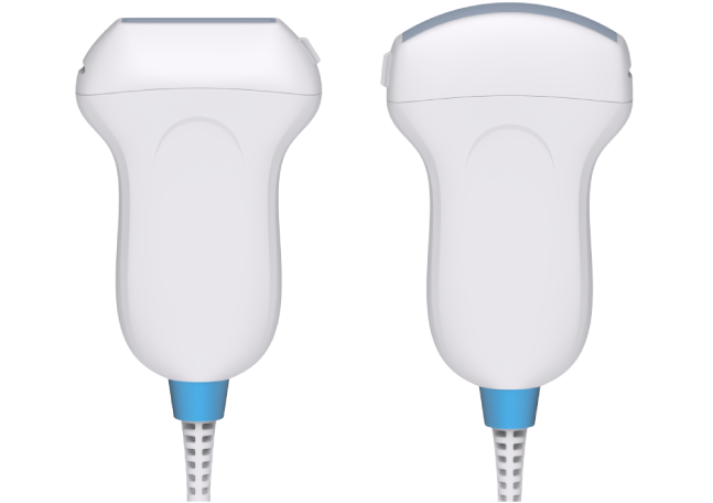
Il MicrUs Pro è un ecografo palmare che fornisce immagini chiare e ad alta definizione con il vostro smartphone, tablet o PC (Windows/Android).

SmartUs
ECOGRAFO PORTATILE HIGH-END
Ecografo portatile con tecnologia Doppler Continuo. Leggero e compatto, garantisce affidabilità, precisione e risultati rapidi per offrire la migliore qualità nell’assistenza sanitaria.

ArtUs
ECOGRAFO PORTATILE HIGH-END
L’ecografo scelto dalle migliori università e dai professionisti sanitari che cercano prestazioni superiori, elevato frame-rate, compattezza e un’ampia gamma di sonde multifrequenza.
